The Sunday News
Kimberly Nyathi
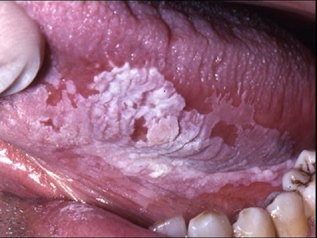
TO understand fully the most common locations of this lesion below is a description of what the term oral mucosa means.
Oral mucosa is all tissues except the teeth themselves in the oral cavity including the gums (gingiva,) mucous membrane covering the cheeks (buccal mucosa), the tongue and its taste buds (papilla) floor of the mouth under the tongue (oral diaphragm) and the roof of the mouth (the palate).
Leukoplakia is a white, firmly attached plaque like lesion found on any of the above mentioned areas of the oral cavity, it cannot be removed and the surrounding oral mucosa is not red or inflamed (hyperemic). It may disappear and reappear on its own.
Causes
(1) Very often the cause is unknown.
(2) Smoking increases the risk of leukoplakia incidence.
(3) Excessive alcohol consumption
(4) Chronic trauma: caused but dental appliances and prosthesis or even bad habits like tongue biting and cheek chewing.
(5) Infection: HPV-Human Papilloma Virus, HIV:Human Immunodeficiency Virus, Candidiasis (oral thrush).
Types
(1) Simple Leukoplakia: flat and usually at the angle of the mouth, usually it is painless.
(2) Verucous Leukoplakia: Above the level of the surrounding mucosa and it is considered premalignant (a lesion that may develop into cancer if left untreated, mostly in males above the age of 30).
(3) Hperkeratotic Verrucous (Complex) Leukoplakia: This type usually occurs on the tongue, usually accompanied with pain and a burning sensation.
(4) Leukoplakia combined with an Erosion (shallow depression the the mucosa — 1 layer deep): Also common in patients above 30, it is also usually painful and accompanied with a burning sensation.
(5) Hairy Leukoplakia: This type is usually associated with patients who are immuno-compromised, HIV positive, Aids, EBV infection (Epstein Barr Virus) and patients with Infectious mononucleosis.
Diagnosis
— Usually objective and subjective examination is adequate: The dentist will ask a few questions about the history of the lesion, some of your habits, and will also visually examine your oral cavity and try to lift or remove the leukoplakia
— A biopsy may be necessary: The dentist may need to cut off the affected mucosa and send it to a laboratory for microscopic examination to determine if it is infectious, cancer causing and to also help decide what kind of treatment is needed.
Treatment
— Depends primarily on the cause, the underlying or causal issue must be addressed first.
— Some analgesics (painkillers) can be prescribed to reduce the discomfort).
— Professional oral hygiene (scaling and polishing) may also be recommended to ensure that no other infection occurs.
— Some topical (cream) antiviral may be used if the cause is infectious.
— If the cause is a dental appliance or prosthesis a new one may need to be fabricated and the old one must not be used.
— The dentist will also place emphasis in the risks of smoking.
The oral mucosa is a mirror of a person’s general health, if you notice any changes in colour, consistency and texture of any of your oral mucosa, see your dentist.
Lee-Anne Hall
BSc Physiotheraphy


